Bilateral Fenestral Otosclerosis
Findings:
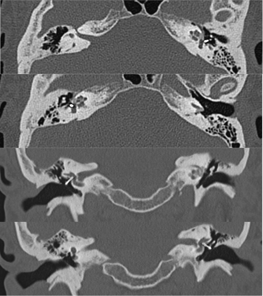
Axial and coronal temporal bone CT images demonstrate bilateral metallic stapes prostheses in normal position. Patchy relatively extensive zones of demineralization are present along the bilateral anterior oval window margins.
Discussion:
When the provided history is conductive or mixed sensorineural hearing loss for temporal bone CT, careful attention to the anterior oval window margins should be paid since this is where otosclerotic plaques form. This is typically much more sublte than the more common inflammatory opacification of mastoid air cells and/or middle ear cavity that can be a distracting or seomtimes incidental finding. Otosclerosis is a misnomer since the imaging findings are lucent (spongiotic) rather than sclerotic, therefore some prefer the term otospongiosis. Additional cases and discussion of otosclerosis:
-otosclerosis w stapes prosthesis
-otosclerosis w obliterative disease
-otosclerosis
-otosclerosis2
BACK TO
MAIN PAGE