DVA and Cavernomas
Findings:
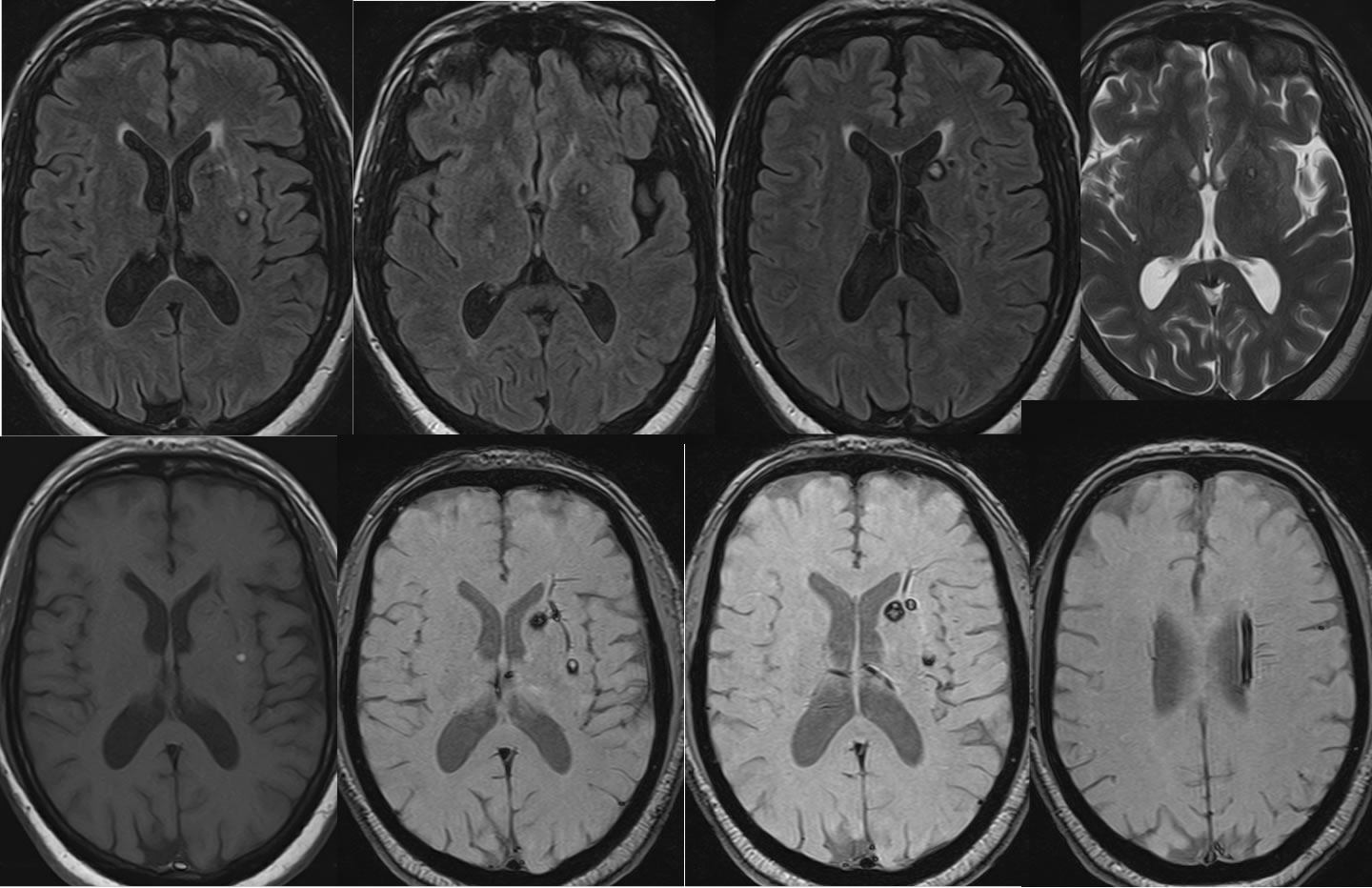
Multiple MR images demonstrate small rounded FLAIR hyperintense foci within the left external capsule, basal ganglia, and caudate nucleus. These lesions are associated with a thick continuous rim of hemosiderin staining confirmed on the susceptibility weighted sequence. One lesion in the left external capsule demonstrates a central zone of T1 hyperintensity. A tubular hypointense structure with branching pattern is also seen to course along the left corona radiata and left basal ganglia.
Differential Diagnosis:
Rounded hemorrhagic lesions have a differential diagnosis of benign vascular findings such as cavernomas in the absence of surrounding edema. A thick continuous hemosiderin rim also indicates probable cavernoma. If edema were present and no thick hemosiderin rims were seen, hemorrhagic metastases should be considered. However, the superimposed developmental venous anomaly also confirms that these represent cavernomas since both may coexist.
Discussion:
Additional examples and discussion of cavernomas:
-Cavernoma1
-Cavernoma2
-Cavernoma3
-Cavernoma4
-cavernoma pontine w acute hemorrhage
BACK TO
MAIN PAGE
