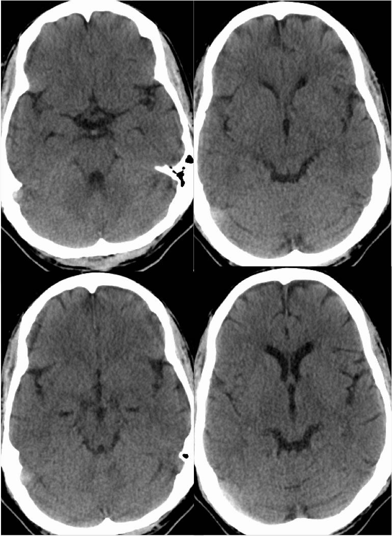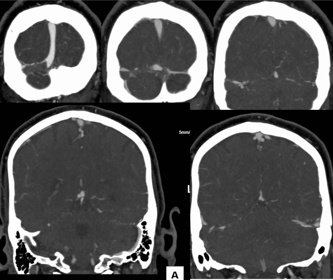

Right Transverse and Sigmoid Sinus Thrombosis
Findings:
Noncontrast CT images demonstrate asymmetric hyperdensity of the right transverse sinus. CT venogram images demonstrate a nonenhancing filling defect that appears occlusive within the right transverse and sigmoid sinuses.
Differential Diagnosis:
The presence of a nonenhancing filling defect within a dural sinus that is occlusive and elongated indicates the presence of thrombosis. Rounded filling defects in the dural sinuses are typically related to arachnoid granulations. There is some overlap in appearance of nonocclusive thrombus and arachnoid granulations when they have an irregular appearannce, but other MR sequences should be helpful to distinguish these. Part of the search pattern on noncontrast CT should include evaluation vascular density. If there is symmetric hyperdensity of arterial and venous structures, this is typically related to hemoconcentration or recent IV contrast administration. An asymmetric hyperdensity on noncontrast CT within dural sinuses or arterial structures should raise the possibility of thrombosis.
Discussion:
Additional discussion and examples of dural sinus thrombosis:
-Superior sagittal
sinus thrombosis
-Superior sagittal
sinus thrombosis2
-Dural sinus thrombosis with hemorrhagic venous infarcts
-Straight sinus
thrombosis
BACK TO
MAIN PAGE


