Osteomyelitis
Findings:
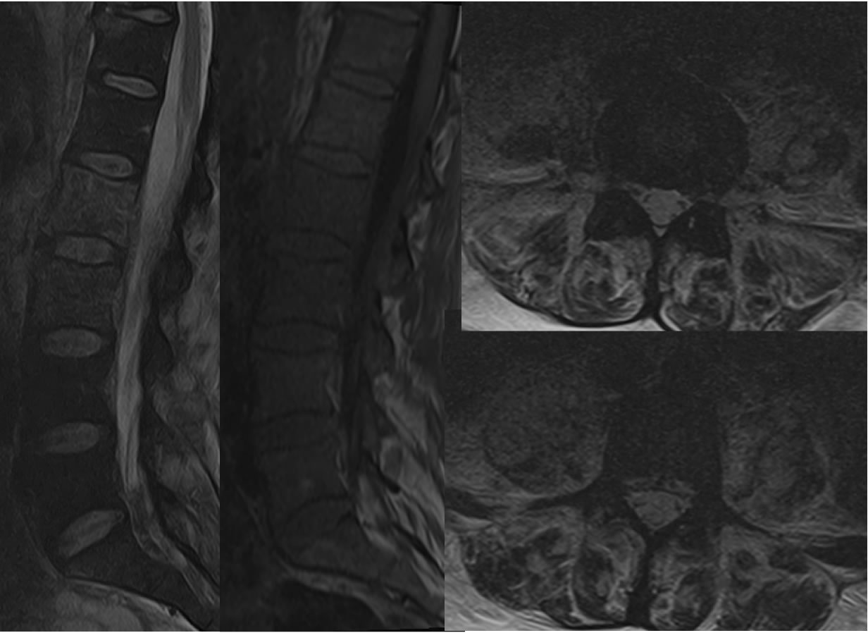
Multiple MR images show hazy abnormal signal involving the L2 and L3 vertebral bodies with normal signal of the L2-3 disc space and adjacent discs. The axial T2 images demonstrate extensive irregular T2 hyperintensities of the psoas muscles with several small fluid collections at least on the left.
Discussion/Differential Diagnosis:
It is unusual to see osteomyelitis involving two consecutive vertebrae without involvement of the intervening disc space, but may conceivable occur from adjacent infection. TB is known to more preferentially involve the spine than discs, but the organism responsible for this case was S. aureus. Other cases of discitis-osteomyelitis and discussion:
BACK TO
MAIN PAGE