Pilocytic Astrocytoma
Findings:
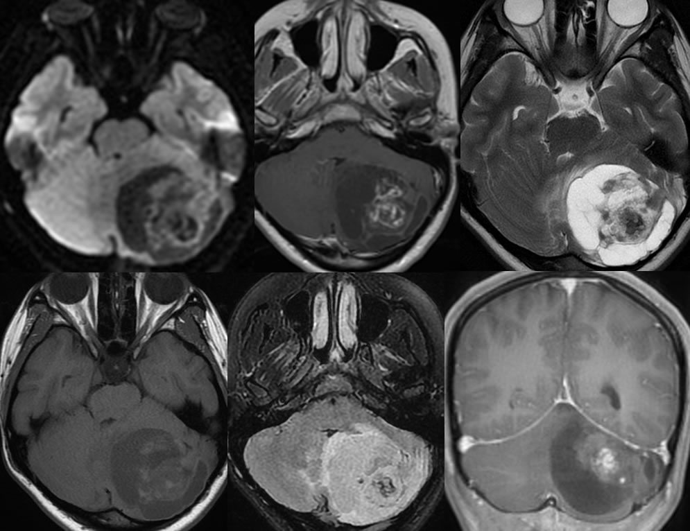
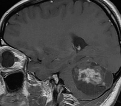
A partially cystic, partially solid mass is centered in the left cerebellar hemisphere. Multiple septations are present within the cystic portion of the lesion. Within the solid portion of the mass, curvilinear and nodular enhancing components are present. Extensive susceptibility within the solid portions consistent with hemorrhage. There is relatively mild surrounding vasogenic edema. Moderate mass effect on the fourth ventricle and brainstem is present. Findings of obstructive hydrocephalus are not included on these images.
Differential Diagnosis:
While there are many possible types of posterior fossa neoplasms, pilocytic astrocytoma, hemangioblastoma, and metastasis are the most likely to show a complex cystic and solid morphology.
Discussion:
Pilocytic astrocytomas are less common in adults than children, representing less than 10% of adult astrocytomas and up to 30% of pediatric astrocytomas. They are most commonly located in the cerebellum, but may also occur in the hypothalamic region or cerebral hemispheres. Less commonly, these lesions are intraventricular or involve the brainstem.
CT and MR imaging demonstrates a complex cystic mass with a mural nodule and minimal surrounding edema. The cyst wall does not typically enhance. The cystic components are typically larger in the cerebellum than when seen in the cerebrum. The cyst fluid typically demonstrates attenuation and signal close to CSF. Calcification is uncommon.
Clinically, these are slow-growing lesions that may present with seizures or other symptoms related to location. With total resection, cure rate approaches 100%. Radiation therapy may be used after subtotal resection, with an overall 85% fysr. Higher grade malignant degeneration may occur in adults and/or after radiation. The histologic analysis will show GFAP positivity and Rosenthal fibers.
For additional discussion, please refer to these cases:
This case was prepared with the assistance of Joshua Hall, UC undergraduate
-Astrocytoma
-pilocytic astrocytoma
-grade 2 astrocytoma
-anaplastic astrocytoma
-anaplastic astrocytoma2
-grade 3 astrocytoma
-medulla astrocytoma