Tumefactive Demyelination
Findings:
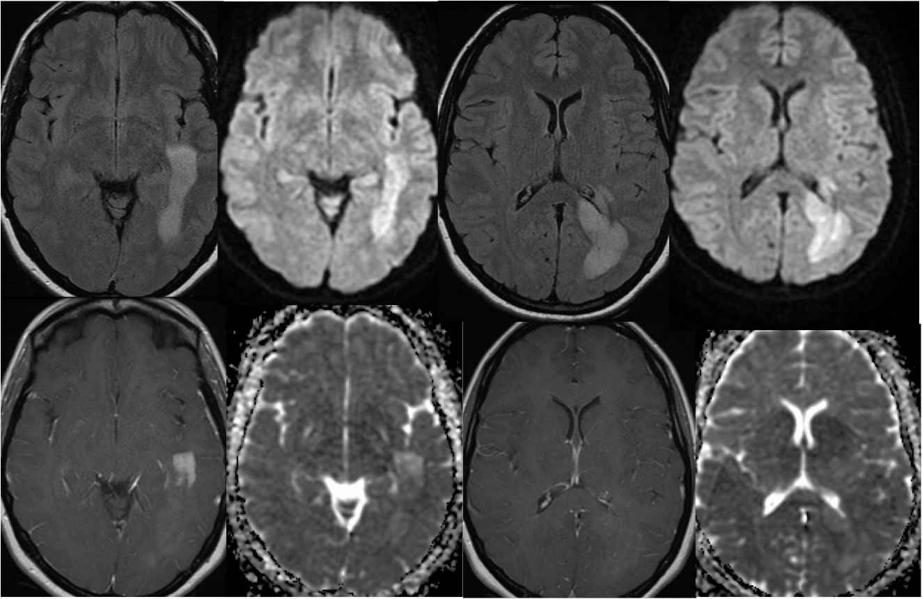
Multiple MR images demonstrate a confluent zone of FLAIR signal alteration in the left temporal lobe and periatrial region with no significant mass effect. Mixed diffusion signal is seen. The process demonstrates patchy enhancement after gadolinium administration, which is most prominent and nodular along the anterior aspect where the enhancement is limited to the leading edge of the signal abnormality.
Discussion/Differential Diagnosis:
The differential diagnosis for an intraaxial enhancing lesion is broad. The typical differential diagnosis includes metastatic disease, glioblastoma or other high grade glial neoplasm, subacute infarct, active MS, or abscess. The lack of mass effect or edema would make mets, GBM, and abscess very unlikely. An enhancing leading edge is typical for demyelination and would not be seen with subacute infarct. Additional cases and discussion of MS and MS like processes:
-optic
neuritis due to MS
-active MS +DWI no enhancement
-Multiple Sclerosis, spinal cord with active lesion
-Susac Syndrome
-tumefactive demyelination
-tumefactive demyelination 2
-neuromyelitis optica
BACK TO
MAIN PAGE