OMU pattern obstructive sinusitis, odontogenic
Findings:
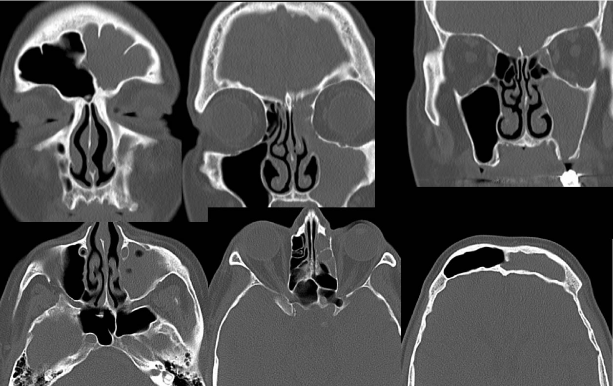
Multiple bone reconstructed CT sinus images in coronal and axial planes demonstrate complete opacification of the left frontal and ethmoid sinuses as well as subtotal opacification of the left maxillary sinus. The walls of the left maxillary sinus are slightly thickened and sclerotic. A defect is present in the floor of the left maxillary sinus on one of the coronal images. The included right sided paranasal sinuses are clear.
Discussion/Differential Diagnosis:
The maxillary ridges are commonly overlooked on sinus CT, while it is quite common for a periapical dental granuloma to erode through the floor of the sinus and cause a more diffuse sinus infection. If no one notes this defect, FESS will be unlikely to cure the inflammatory disease. When there is unilateral sinus opacification, an underlying cause should be sought. The presence of a sclerotic thickened wall indicates chronic inflammatory disease, with superinmposed defect potentially raising the possibility of a chronic fungal infection. Smoothly remodeled and expanded walls indicate a benign process such as mucocele, uncommon in the maxillary sinus. The osteomeatal complex is the common drainage pathway for the maxillary and ethmoid sinuses, and if obstructed by inflammatory disease, polyp, or other mass, these sinuses often become completely opacified. Additional cases and discussion of sinus inflammatory disease:
-Sphenoid Polyps
-frontal sinus mucocele
-maxillary sinus mucocele
-antrochoanal polyp
-invasive fungal sinusitis
-Chronic maxillary sinusitis, odontogenic origin
-odontogenic abscess
BACK TO
MAIN PAGE