Complex Basilar Artery Aneurysms, severe stenosis basilar and PCA
Findings:
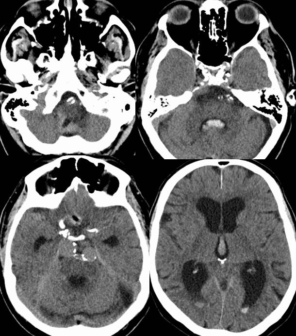
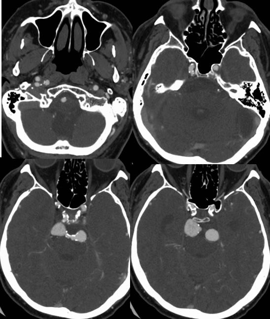
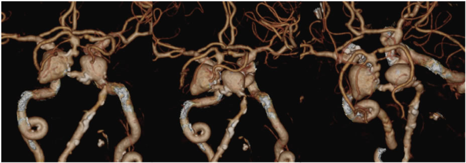
Axial noncontrast CT images demonstrate a dilated ventricular system with a small amount of intraventricular hemorrhage. Extensive vascular calcifications are present. Source images from CTA demonstrate two large lobulated aneurysmal projections of the basilar artery. The volume rendered images also demonstrate a severe stenosis of the basilar artery just proximal to the aneurysm and additional significant stenosis of the left P1 segment.
Differential diagnosis:
None other than aneurysm really applies. Prepontine extraaxial soft tissue density masses may also be seen with meningioma, schwannoma, chordoma, and metastases.
Discussion:
Discussion of aneurysms and associated complications is found on several other cases on this site including these:
BACK TO
MAIN PAGE