Meningoencephalitis with Empyemas
Findings:
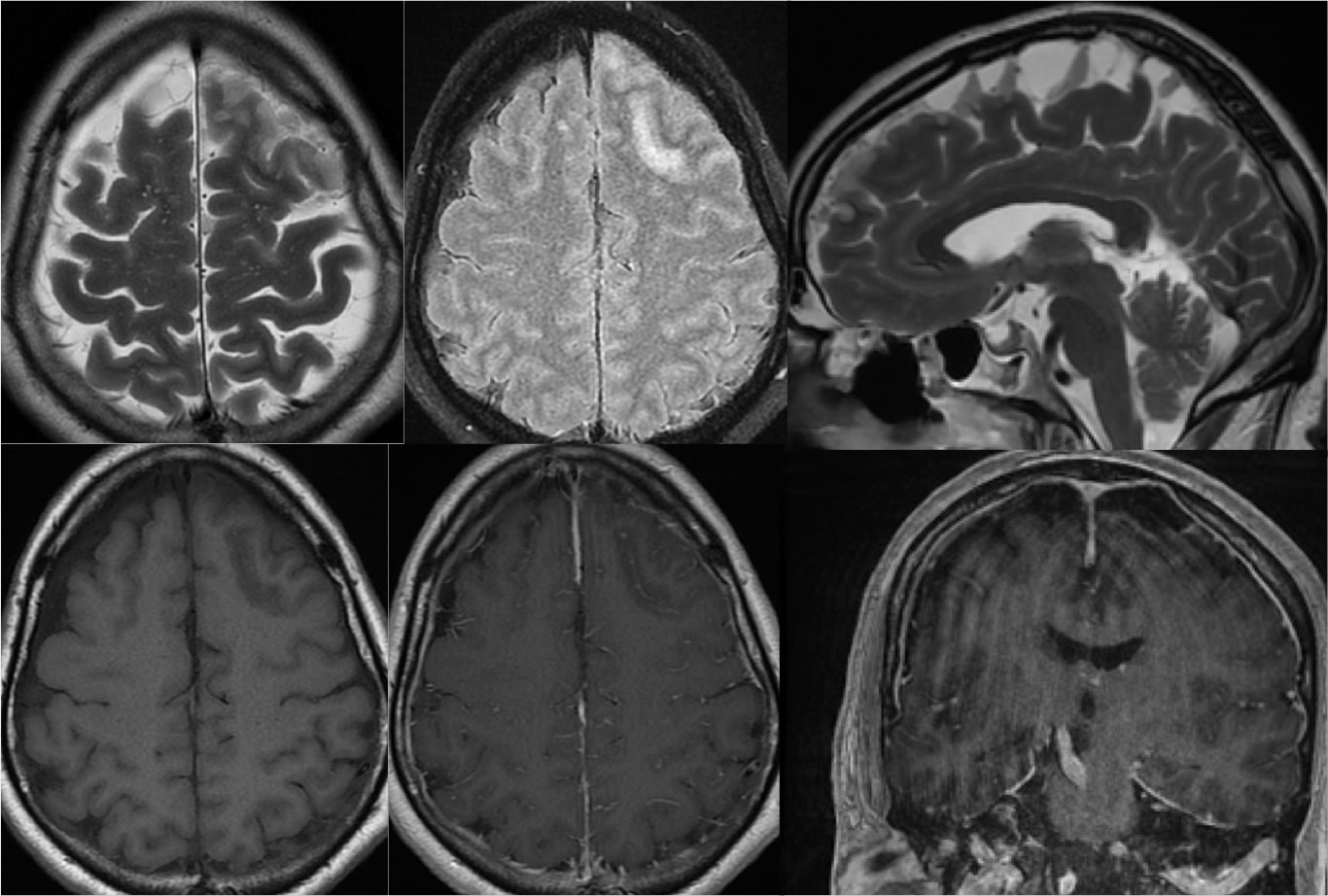
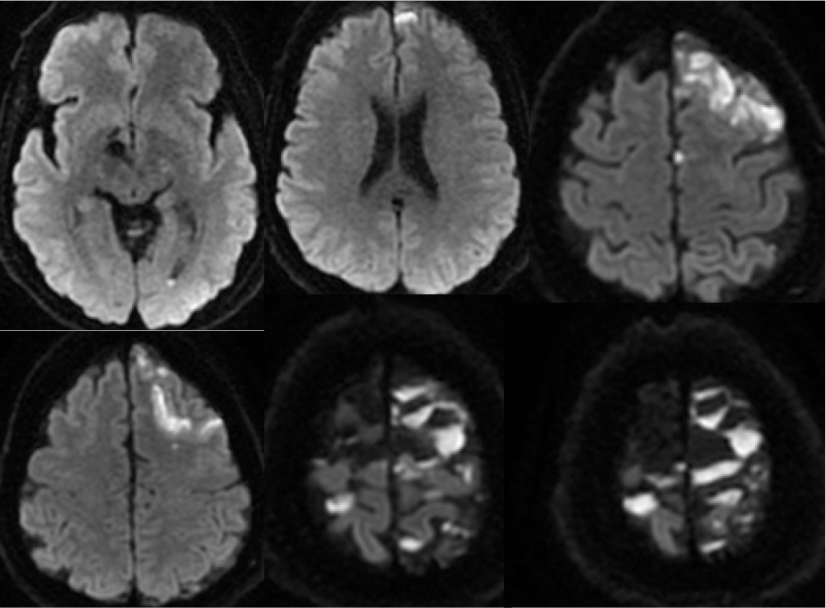
The axial T2 weighted image demonstrates abnormal signal intensity in a patchy distribution of the left frontal cortex. The axial FLAIR image demonstrates the same findings as well as abnormal hypointensity of the white matter underlying the cortical signal changes. There is abnormal FLAIR hyperintensity within the subarachnoid spaces. Subtle leptomeningeal enhancement in this region is seen on the post contrast images. The sagittal T2 and coronal T1 post contrast images demonstrate loculated fluid collections near midline at the vertex with fluid-fluid levels and irregular peripheral enhancement. The diffusion weighted imaging demonstrates restricted diffusion within the fluid collections over the vertex, as well as zones of restricted diffusion corresponding to the T2 cortical hyperintensities. There is also a small focus of restricted diffusion within the left occipital horn.
Discussion/Differential Diagnosis:
The presence of restricted diffusion within extraaxial spaces is characteristic for infection, as long as the difference between a subdural hematoma and a subdural empyema is recognized. Other cases and discussion of intracranial infection:
BACK TO
MAIN PAGE