Glioblastoma Multiforme
Findings:
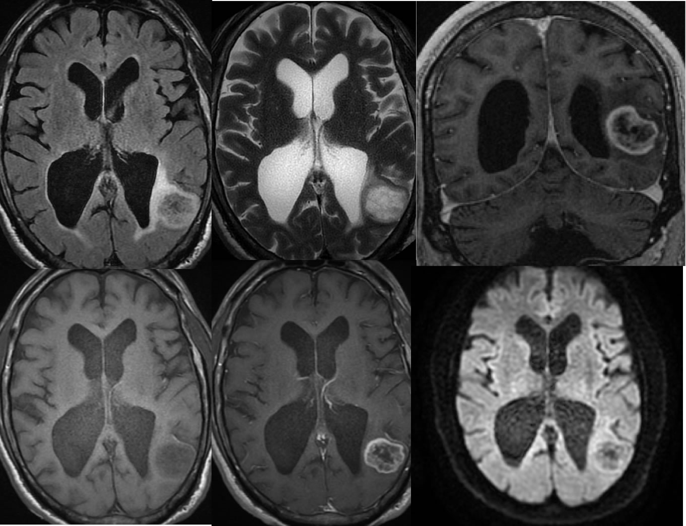
Multiple MR images demonstrate a well circumscribed mass in the left parietal lobe which demonstrates irregular peripheral enhancement, minimal surrounding FLAIR signal alteration, no significant diffusion restriction, and very little mass effect for the size of the lesion. The lateral ventricles are asymmetrically dilated with slight effacement of the left lateral ventricle by the mass.
Discussion/Differential Diagnosis:
GBM may have variable appearance. GBM are known to be high grade aggressive malignancies, but occasionally may appear better circumscribed and are associated with less mass effect than expected for the size of the enhancing mass. Multiple other cases and discussions of GBM are found on this site including below:
BACK TO
MAIN PAGE