Grade 3 Anaplastic Astrocytoma
Findings:
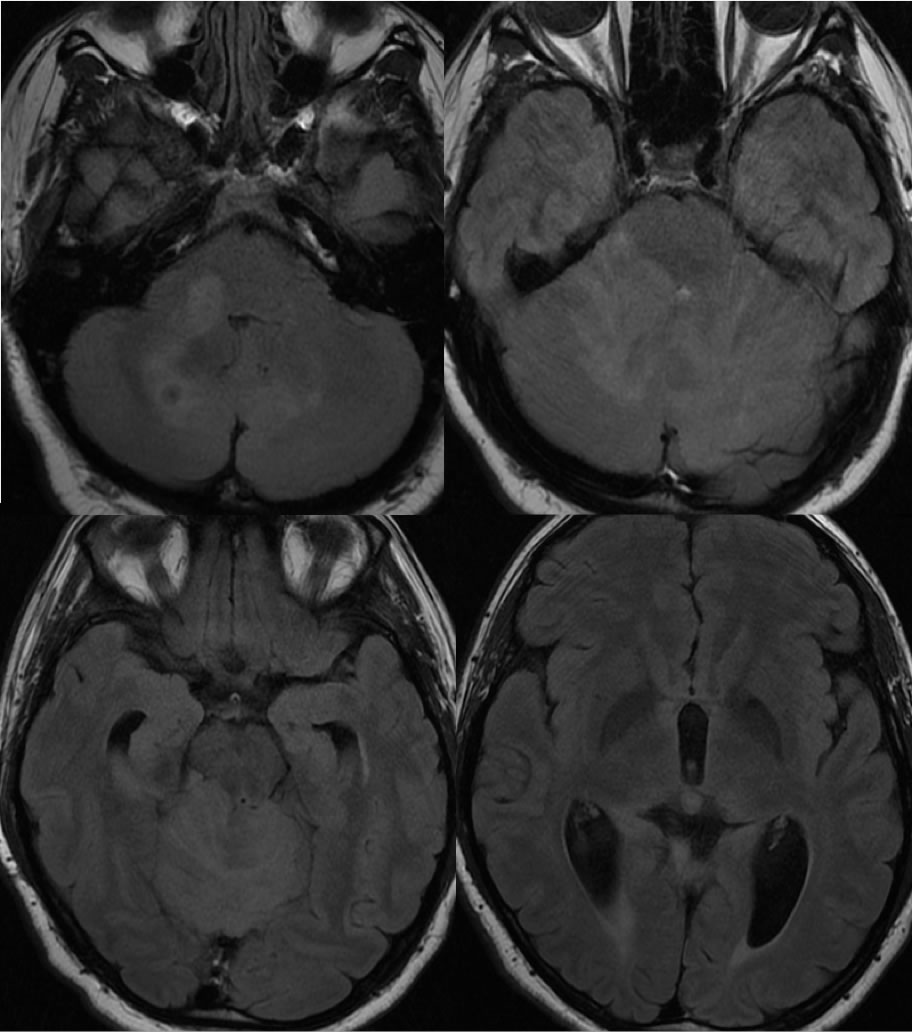
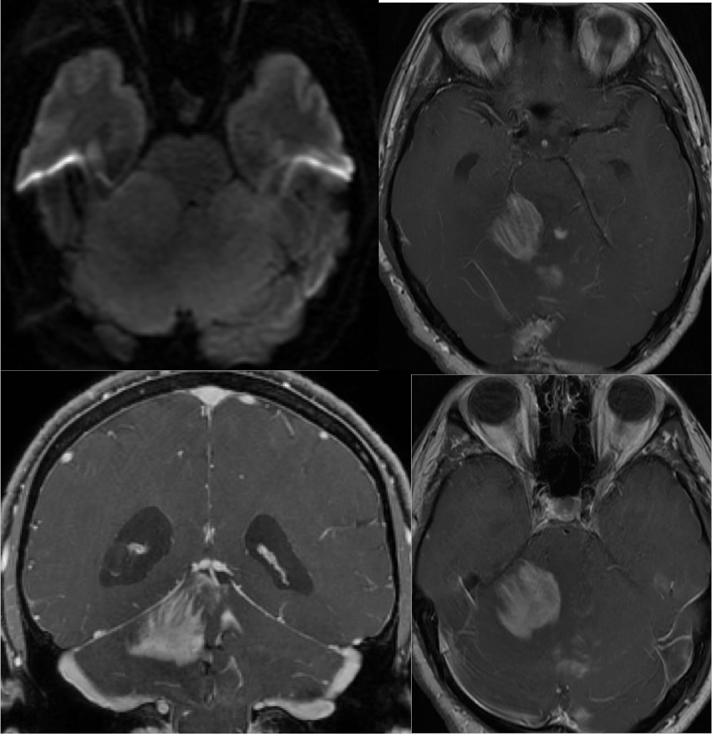
Axial FLAIR images demonstrate irregular patchy signal abnormalities with mass effect involving the bilateral cerebellum. The fourth ventricle is moderately effaced. The lateral and third ventricles are mildly dilated due to obstructive hydrocephalus. No diffusion restriction is evident. The axial and coronal post contrast images demonstrate multifocal poorly defined enhancement in the cerebellum, with some streaklike and gyriform morphology, while other zones are nodular.
Discussion/Differential Diagnosis:
The apparent multifocal nature of this process should raise the possiblity of metastases, but the configuration of enhancing gyriform lesions and nonenhancing lesions with mass effect would be unlikely to represent metastases. Other possibilities include encephalitis (unusual to have absent DWI hyperintensity) and PML/IRIS (no known rx for demyelinating disease). Glial neoplasms including anaplastic astrocytoma should be considered first for this appearance, and this is not anectodally an unusual appearance for cerebellar intermediate to high grade glioma. Other cases and discussion of primary glial neoplasms:
BACK TO
MAIN PAGE