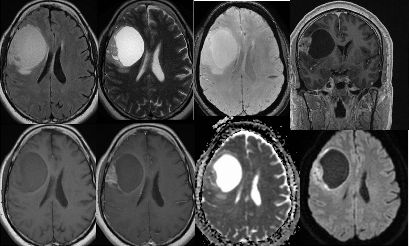
Grade 3 Astrocytoma
Findings:
A complex cystic mass is present in the right frontal lobe with an irregular zone of nodular enhancement along the lateral margin. Thin rim enhancement is seen more medially. The cystic portion does not demonstrate restricted diffusion. Decreased ADC values are seen within the enhancing nodular portion of the mass. There is very little surrounding edema for the size of the lesion, with mild to moderate mass effect. The nodular enhancing portion is inseparable from the dura laterally.
Differential Diagnosis:
The differential diagnosis for a rim enhancing intraaxial lesion is relatively broad, including glioma and metastasis. A greater degree of surrounding vasogenic edema would be expected for metastasis. The absence of central restricted diffusion is not compatible with abscess, and again the surrounding vasogenic edema would be more prominent. The relatively little surrounding vasogenic edema and mass effect for the size of the lesion and other characteristics are compatible with an intermediate to high-grade primary glial neoplasm.
Discussion:
Additional discussion and examples of gliomas:
-GBM
-GBM corpus callosum
-GBM w subependymal
-GBM intraventricular recurrence
-GBM spine w CSF dissemination
-recurrent GBM w uncal herniation
-recurrent GBM, corpus callosum
-GBM left frontal
-glioma/gliomatosis
cerebri
-oligodendroglioma
-pontine glioma
-Gliosarcoma
-Astrocytoma
-pilocytic astrocytoma
-pilocytic astrocytoma2
-grade 2 astrocytoma
-anaplastic astrocytoma
-anaplastic astrocytoma2
-grade 3 astrocytoma
-medulla astrocytoma
-anaplastic oligodendroglioma
-spinal cord astrocytoma
-spinal cord astrocytoma pilocytic
BACK TO
MAIN PAGE

